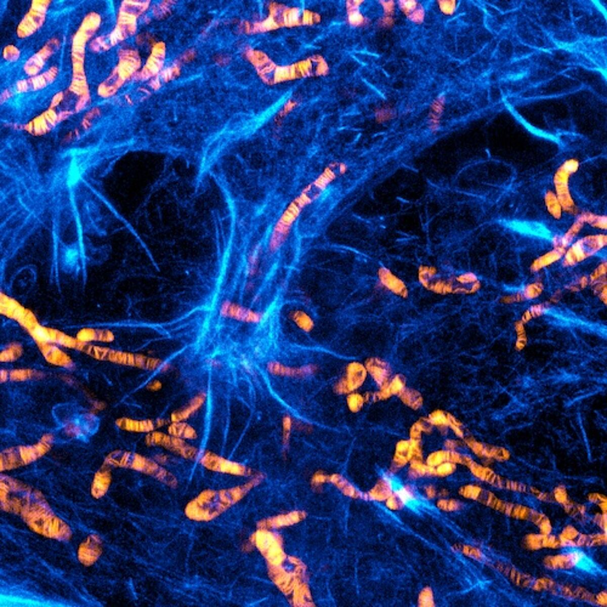
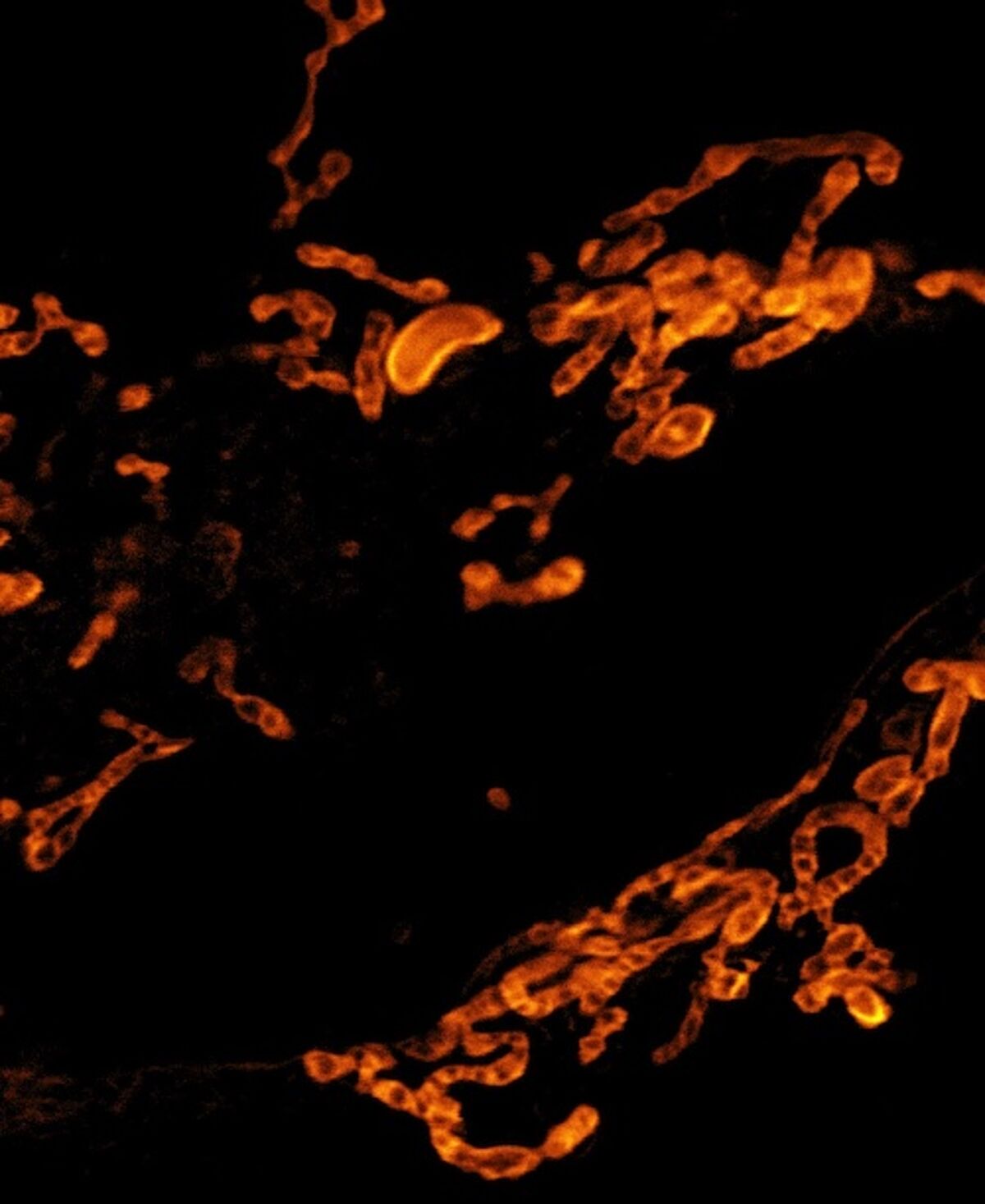
PKmito™ probes are bright and photostable fluorescent probes for gentle imaging of mitochondria in live cells. PKmito™ probes consist of cyanine derivatives bearing intramolecular cyclooctatetraene (COT) substituents. The presence of intramolecular COT substituents very efficiently reduces the production of singlet oxygen. Singlet oxygen is a very reactive species produced by fluorophores and it is responsible for rapid cellular damage, leading to cell death. PKmito™ probes allow very gentle imaging of mitochondria in live cells and tissues, preventing singlet oxygen damage and phototoxicity.
Available PKmito probes:
- PKmito RED
- PKmito ORANGE
- PKmito ORANGE FX (for fixed cells)
- PKmito DEEP RED
Advantages
- Very low phototoxicity, allows recording of time lapse STED movies (ca 20 frames) without damaging or changing mitochondria morphology
- Long-term live cell imaging with confocal or widefield microscopes
- Ideal for STED, staining the inner mitochondrial membrane and revealing mitochondrial cristae impossible to see with conventional microscopes
- Very easy to use, also in combination with SiR and SPY probes
PKmito RED
Excitation: 549 nm
Emission: 569 nm (Cy3 channel)
Needs 660 nm STED depletion line, STED-compatible but not optimal.
PKmito ORANGE
Excitation: 591 nm
Emission: 608 nm (custom channel with 580-590 nm excitation, 600 and 700 nm emission)
Works with 775nm nm STED depletion line, highly suited for STED.
PKmito ORANGE FX
PKmito ORANGE FX is based on the original PKmito ORANGE probe and has been optimized for imaging of mitochondria in formaldehyde and glutaraldehyde fixed cells. The unique and unmatched feature of PKmito Orange FX is its ability to be retained nearly quantitatively after aldehyde fixation of stained cells.
Excitation: 584 nm
Emission: 604 nm
Works with 775nm nm STED depletion line, highly suited for STED.

PKmito DEEP RED
Excitation: 644 nm
Emission: 670 nm (Cy5 channel)
Works with 775nm nm STED depletion line, well suited for STED.
Frequently asked questions
Do PKmmito probes work in fixed cells? No, PKmito probes are intended for live cell imaging only.
Do PKmmito probes work in plant cells? This hasn't been tested yet. Please contact us if you are interested in testing PKmito probes in plant cells.
Do PKmmito probes work in tissue? Yes.
Are PKmito probes sensitive to mitochondrial membrane potentials? Yes, mitochondrial deploarisation (e.g. using FCCP) leads to a 50% decrease of fluorescence signal within 30 minutes.


