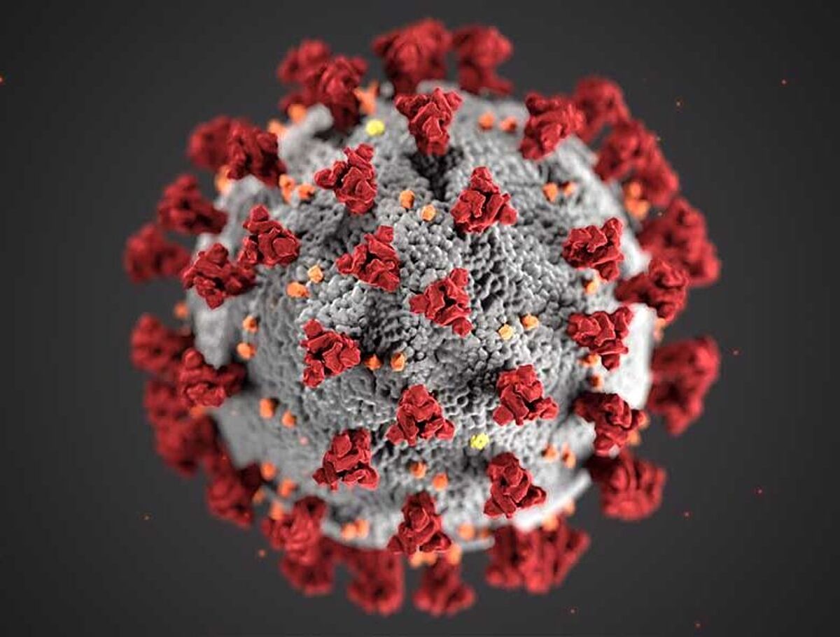
As of May 2021, over 153 million people have been diagnosed with COVID-19 and over 3.2 million have died from the virus globally (https://coronavirus.jhu.edu/map.html). Scientists world-wide continue to investigate how SARS-CoV-2 infects our bodies to develop better diagnostics, treatments, and vaccines.
While COVID-19 is primarily a respiratory disease, it affects multiple organ systems—including the central nervous system (CNS), which has presented as impaired consciousness and headaches among others (Mao et al., 2020). However, there is no consensus on the consequences of CNS infections and there are several unknowns surrounding the frequency of neuroinvasion. Unmasking the various ways patients are affected by SARS-CoV-2 is crucial to treating patients as well as understanding long-term consequences.
There are several challenges when studying neurological disease in COVID-19 patients, including
- Only a subset of COVID-19 patients show neuroinvasion, limiting sample size and access to samples,
- A lack of technology to sample CNS tissues directly, and
- The ability to distinguish between direct neuroinvasion and systemic viremia within the brain.

Song et. al. realized that, to overcome the challenges of studying COVID-19 neuroinvasion, they needed robust, reliable model systems.
To get closer to understanding the underlying SARS-CoV-2 neuropathology, Song et. al. probed the capacity of SARS-CoV-2 to infect the brain using three independent approaches:
- Human brain organoids
- Mice overexpressing human ACE2
- Postmortem COVID-19 patient brain tissues
Here’s a breakdown of what the researchers did with each approach:
| Human brain organoids | Mice overexpressing human ACE2 | Postmortem COVID-19 patient brain tissues |
|---|---|---|
Used human induced pluripotent stem cell lines (hiPSC) to test the infection capacity of SARS-CoV-2 in CNS tissue. Used single-cell RNA sequencing (RNA-seq) to uncover transcriptional changes caused by SARS-CoV-2 infection of neurons. | Used transcriptional profiling along with blocking antibody studies to demonstrate ACE2 and SARS-CoV-2 spike protein requirements for neuronal infection. Observed vascular remodeling in infected regions. | Examined sections of three patients who died after suffering from severe COVID-19–related complications. Provided evidence of neuroinvasion by SARS-CoV-2. Identified associations between infection and ischemic infarcts in localized brain regions. |
Human brain organoids
The researchers observed that SARS-CoV-2 can infect cells of neural origin and induce metabolic changes in infected and neighboring neurons, potentially promoting death of nearby cells. Further, the researchers found that blocking cell surface protein ACE2 significantly inhibited SARS-CoV-2 infection.
Here are some highlights from the experiments using human brain organoids:
- Using electron microscopy, the researchers visualized viral particles within the organoid, with discrete regions of high-density virus accumulation and other regions showing viral budding from ER-like structures
- Infection was primarily in neuronal cells with a deep-layer fate (CTIP2-positive, PAX6-negative cells or CTIP2/TBR1 double-positive, Pax6-negative cells), but also upper-layer cortical neurons were susceptible (SATB2-positive, CTIP2-negative cells or SATB2/CTIP2 double-positive cells)
- SARS-CoV-2 induced a unique transcriptional state within neurons compared with ZIKV and compared with neighboring uninfected cells without SARS-CoV-2 transcript
- Infection by SARS-CoV-2 induced a locally hypoxic environment in neuronal regions, manipulating host metabolic programming, potentially creating a resource-restricted environment for cells
- Pretreating organoids with ACE2 antibody compared with isotype control inhibited SARS-CoV-2 and cerebrospinal fluid (CSF) from a patient hospitalized with COVID-19 and acute encephalopathy blocked SARS-CoV-2 infection in organoids.
Mice overexpressing human ACE2
The researchers demonstrated SARS-CoV-2 neuroinvasion in vivo and mapped the distribution of infection throughout the brain as well as the impact on vasculature.
Here are the highlights:
- SARS-CoV-2 was widely present in neural cells throughout the forebrain
- Most brain regions contained a high density of infected cells, except for the cerebellum
- Brain regions with a low density of infected cells included the dentate gyrus, the globus pallidus, cortical layer 4, and the vascular endothelium
- The cortex was nonhomogeneously infected, with infected cells seen in columnar patches and in sensory regions
- SARS-CoV-2 infection coincided with a disruption in the normal vascular topology in the cortex
Postmortem COVID-19 patient brain tissues
By examining brain regions of three patients who died after experiencing severe COVID-19–related complications, the researchers found the presence of ischemic damage and microinfarcts. SARS-CoV-2 infection was detected within the regions of micro–ischemic infarcts, suggesting that neuroinvasion is associated with ischemia and vascular anomalies.
Due to the small number of samples, these results cannot be generalized, but do offer a snapshot of case reports from several patients.
Song et. al. provide evidence for SARS-CoV-2 neuroinvasion and an unexpected consequence of direct infection of neurons. The data also give us insights that SARS-CoV-2 neuroinvasion may cause hypoxic regions and vasculature disturbance, and the disruption of brain vasculature can make vulnerable ischemic infarcts vulnerable to viral invasion.
Importantly, this research highlights the utility of human brain organoids and the importance of using diverse approaches to model human physiology, particularly when studying the brain.
Read the full Article
Song, Eric et al. “Neuroinvasion of SARS-CoV-2 in human and mouse brain.” The Journal of experimental medicine vol. 218,3 (2021): e20202135. doi:10.1084/jem.20202135
Or watch a webinar presentation by first-author Dr. Song
Antibody supplier GeneTex invited Dr. Song to discuss his work during a webinar on 4th May, 2021. But don't worry if you missed it - you can watch a recording of the webinar!
Among the antibodies used in the Song et al. 2021 study were two of GeneTex’s SARS-CoV-2 antibodies (GTX635679 and GTX632604), as well as GeneTex’s widely published HIF-1alpha antibody (GTX127309).
Are you interested in GeneTex antibodies? Try them out without risk! Check out their 10ul for 10 CHF promotion (incl. GTX127309)

