Who says science isn't art?
In June, we hosted an Immunohistochemistry or Immunofluorescence art contest. Every contestant received a 20% discount for their next order of VectaPlex by Vector Laboratories. The main prize awarded to the masterpiece chosen by the LubioScience team was a 100 CHF Galaxus voucher.
Congratulations to our winner!
Congratulations to Dr. Cristina Tocchini on winning our art contest with her stunning image of a heart-shaped hatching C. elegans larva.
Thank you to all participants for the beautiful entries, which can be viewed below.
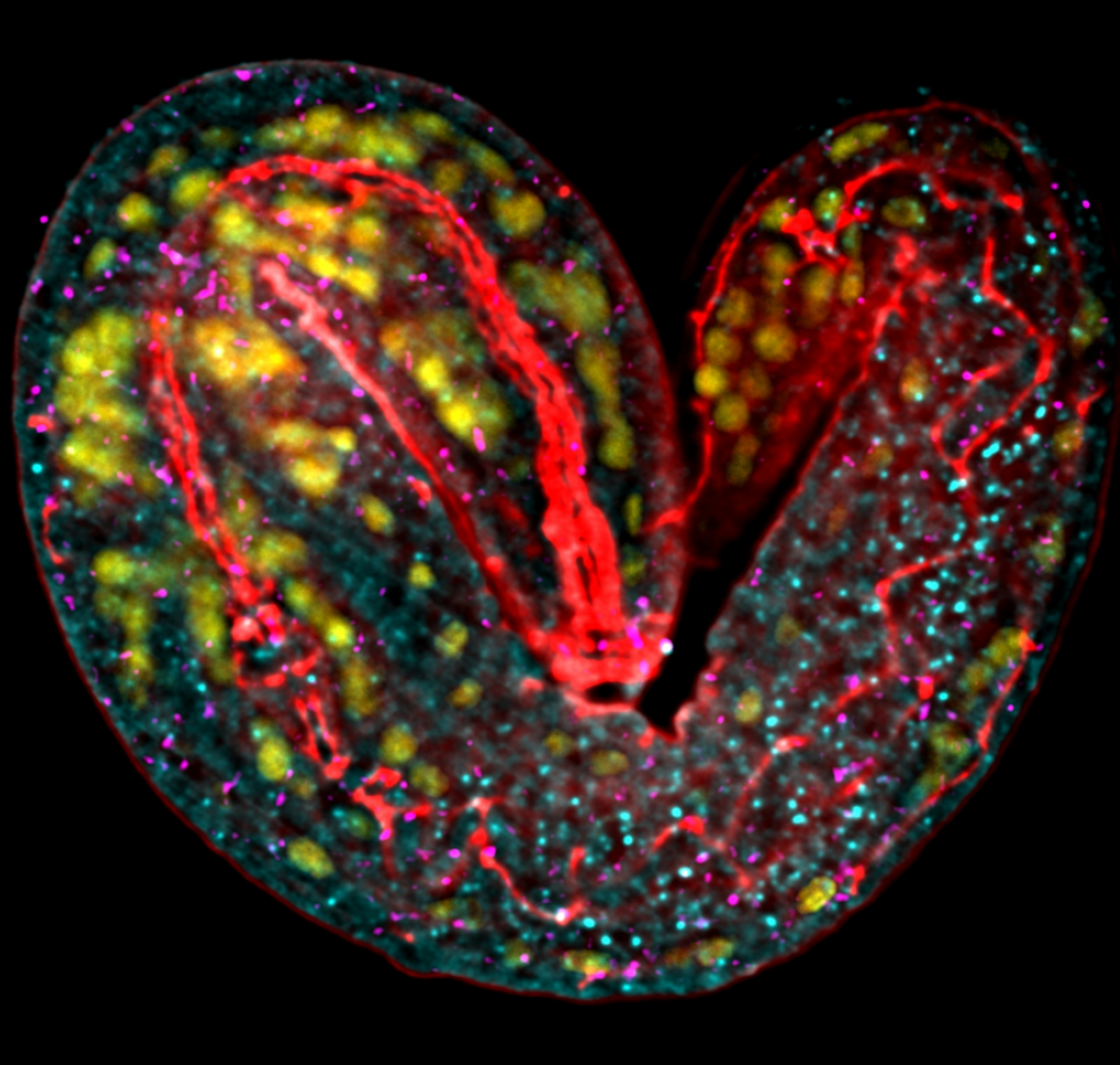
Heart-shaped hatching C. elegans larva by Dr. Cristina Tocchini. Images were acquired with a widefield microscope FEI “MORE” with total internal reflection fluorescence (TIRF), equipped with a Hamamatsu ORCA flash 4.0 cooled sCMOS camera, and a Live Acquisition 2.5 software. Deconvolution has been applied with the Huygens software. Yellow (434 channel): nuclei stained with Hoechst (Thermo Scientific). Red (517 channel): DLG-1 protein stained with antibody PSD95 (7E3) Mouse mAb #36233 (Cell Signaling Technology). Cyan (590 channel): erm-1 mRNA stained with DNA primary probes paired to fluorescently-tagged FLAP probes (ATTO550) (Integrated DNA Technologies - IDT). Magenta (685 channel): spc-1 mRNA stained with DNA primary probes paired to fluorescently-tagged FLAP probes (ATTO647) (Integrated DNA Technologies - IDT).
Art gallery
Christian Zuppinger
University Hospital Bern

hiPSC-derived epicardial progenitor cells immunostained for all-actin (green) and alpha-smooth muscle actin (red)
Anonymous
University of Lausanne

Open and closed valves in collecting lymphatic vessel.
Elisa Rodrigues Sousa
University of Bern

This figure represents a murine prostate stained for different stromal markers. the title of this image is "Unexpected love".
Dario Speziale
Roche

Representative image of an adult mouse kidney cortex that has been processed for 3D immunofluorescence staining and imaged with confocal microscopy (63X oil immersion objective). Epithelial tissue is identified with an E-Cadherin antibody (cyan). Vasculature is stained with CD31 (yellow) and highlights the presence of 4 glomeruli. pH sensitive molecule (magenta) shows areas with pH lower than 6.7, revealing the presence of compartmentalized low/physiological pH in the kidney tissue which is of pharmaceutical interest.
Scale bar is 50 um.
Simone Leoni
University of Bern

Rat cerebellum infected with Tick-Borne Encephalitis Virus (TBEV).
Purkinje cells labeled with Calbindin (green) and TBEV labeled with 4G2-anti-flavivirus antibody (red).
Jeremiah Bernier-Latmani
University of Lausanne

Small intestinal villi harbor telocytes (green, Lgr5-GFP) at their tips that promote blood vessel (red, VEGFR2) stability and function.
In partnership with
Vector Laboratories
Vector Laboratories is recognized as a pioneer and market leader in immunohistochemistry, immunofluorescence, glycobiology, and bioconjugation products.