Imaging of biological structures in living cells without transfections, without phototoxicity, without tedious washing protocols and without unspecific background staining?Spirochrome has developed a whole range of reagents and probes using its innovative silicon-rhodamine and SPY fluorophores to allow imaging in living cells with unprecedented resolution and low background. Spirochome probes are ideal for live imaging and STED/STIM applications.
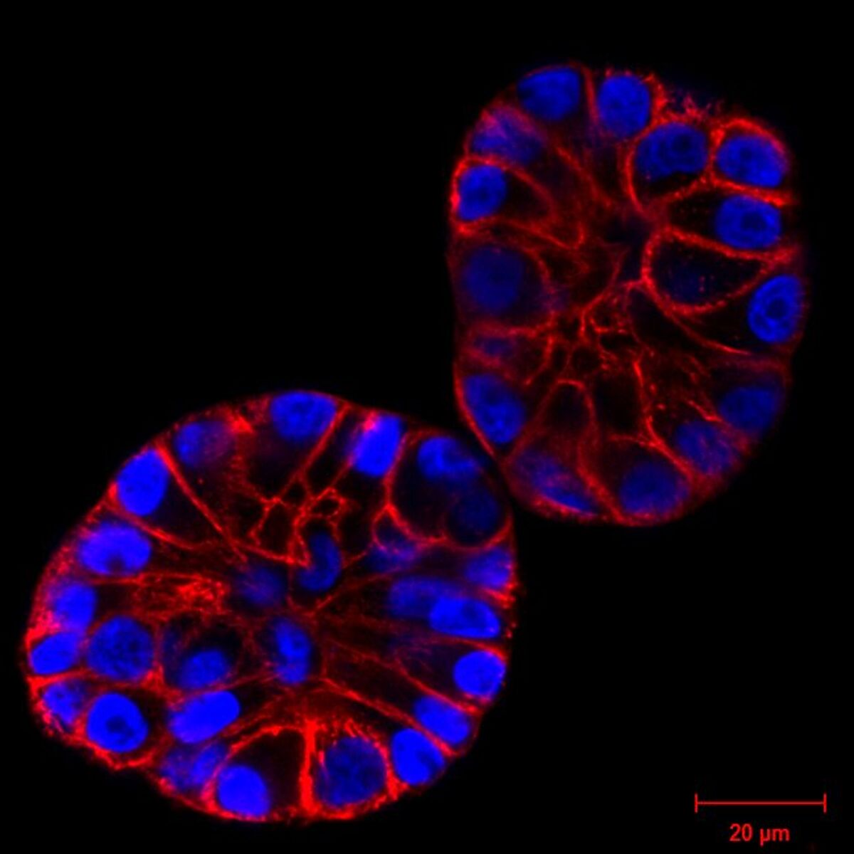
Probes for imaging of cellular structures
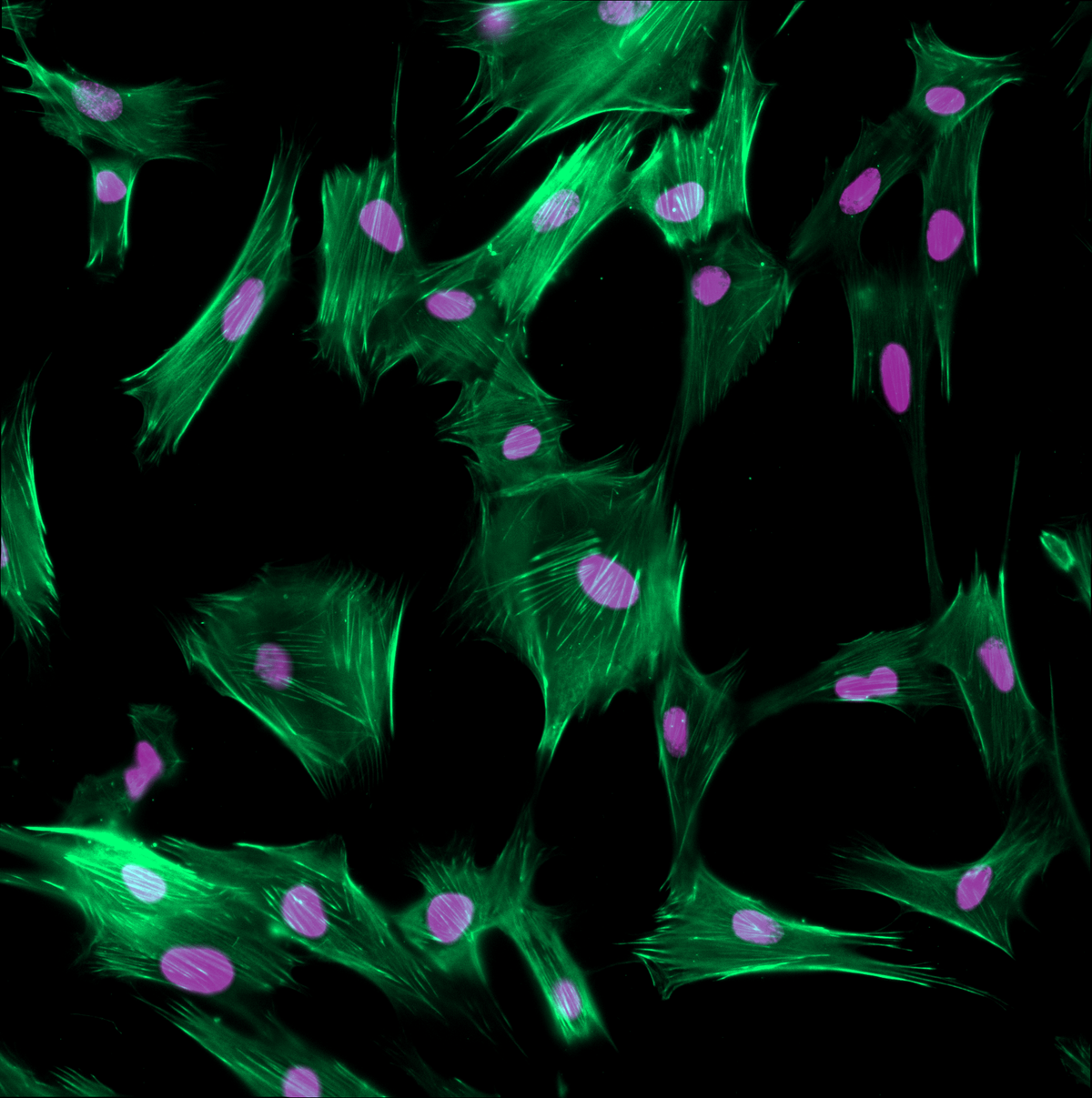
- Cytoskeletal probes (SiR-actin, SPY-actin, SPY-FastAct)
- Microtubular probes (SPY-tubulin, SiR-tubulin)
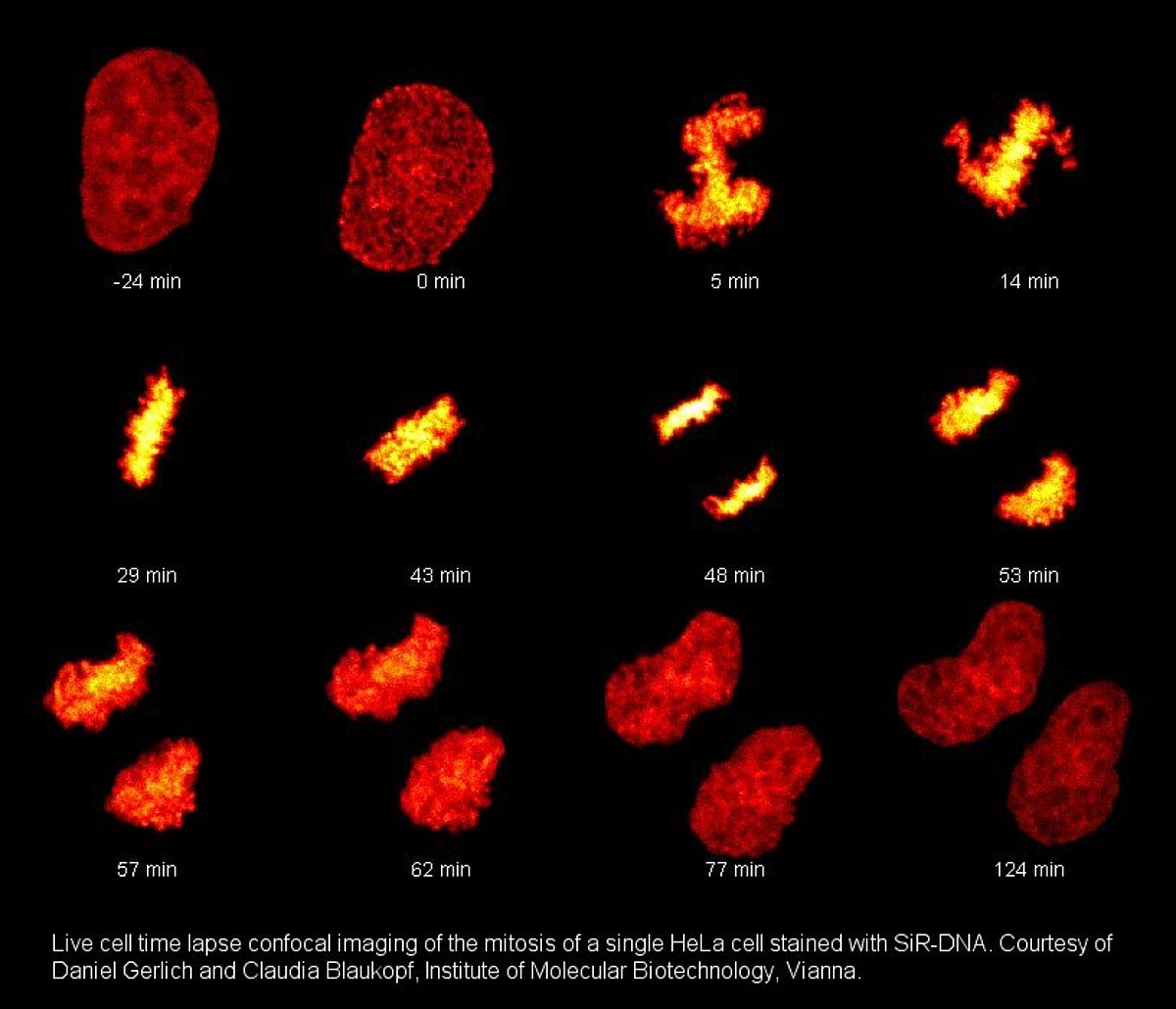
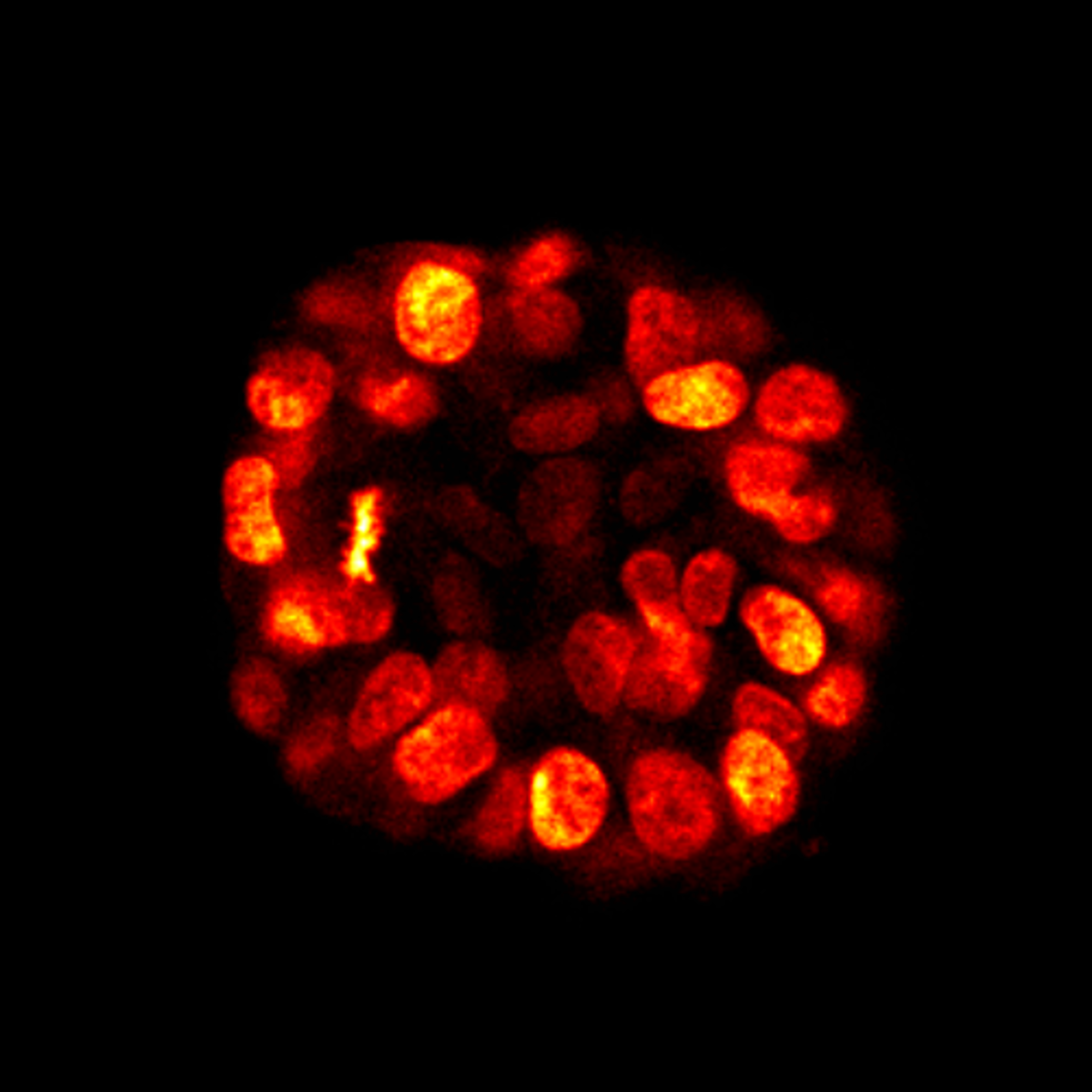
- Nuclear probes (SPY-DNA, SiR-DNA)
- Mechanosensitive membrane probes (Flipper-TR. ER Flipper-TR, Lyso Flipper-TR, Mito Flipper-TR)
- Mitochondrial probes (PKmito RED, ORANGE and DEEP RED)
SPY probes
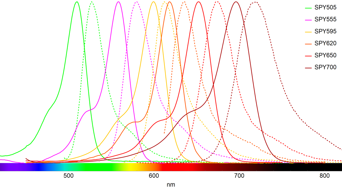
Spirochrome’s SPY™ probes are revolutionary fluorogenic live cell fluorescent probes covering the whole visible spectrum
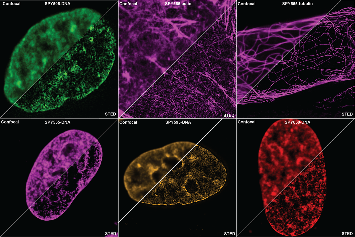
SPY™ probes are live cell, fluorogenic, photostable and STED/SIM compatible. They keep all the nice properties of our SiR-probes but work in different channels such as FITC, TMR or Texas Red. Probes for DNA (nucleus), actin and microtubules are available.
SPY-DNA probes are a better alternative for Hoechst dyes
The are numerous advantages to using SPY-DNA when compared to classical Hoechst, in particular for long term imaging.
- The wavelength used to excite Hoechst (ca 450 nm) is highly phototoxic and induces cell death. SPY650-DNA uses far-red excitation (Cy5 filters or settings) which is much more gentle for the cells.
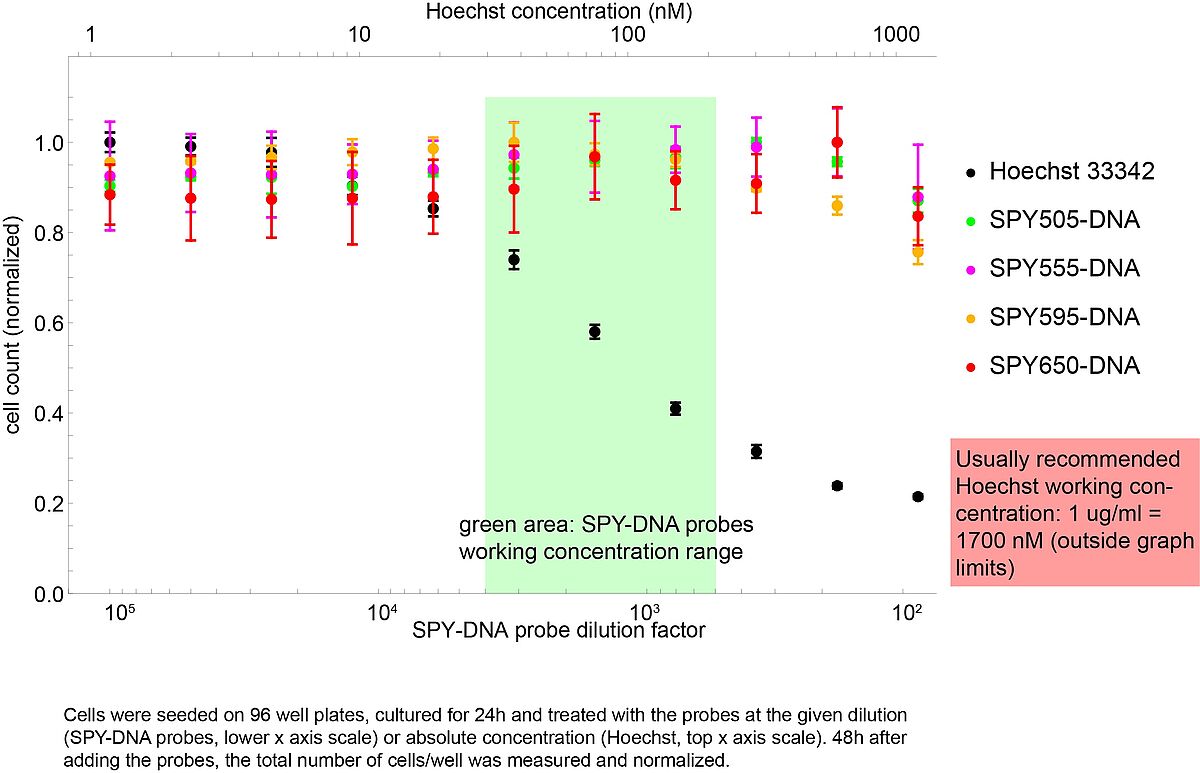
- Hoechst itself has a strong cytotoxicity, even without illuminating the cells. This cytotoxicity strongly increases with the Hoechst concentration used.
Consider the toxicity assay shown on the right. Different dilutions of SPY-DNA probes (green/red/pink/yellow dots) were compared with different concentrations of Hoechst dye (black dots) and cell survival was measured. Toxic effects of Hoechst appear at 50 nM, well below the typical recommended concentration of 1700 nM (1 ug/ml). SPY-DNA probes show no toxicity in the working dilution window (green area).
SPY products
| Cat-No. | Item | Size | Price (CHF) |
|---|---|---|---|
| SC101 | SPY505-DNA | 100 stainings | 330.00 |
| SC201 | SPY555-DNA | 100 stainings | 330.00 |
| SC202 | SPY555-Actin | 100 stainings | 435.00 |
| SC203 | SPY555-Tubulin | 100 stainings | 435.00 |
| SC204 | SPY555-BG | 35 nmol | 330.00 |
| SC301 | SPY595-DNA | 100 stainings | 330.00 |
| SC401 | SPY620-DNA | 100 stainings | 330.00 |
| SC402 | SPY620-Actin | 100 stainings | 435.00 |
| SC404 | SPY620-BG | 35 nmol | 330.00 |
| SC501 | SPY650-DNA | 100 stainings | 330.00 |
| SC503 | SPY650-Tubulin | 100 stainings | 435.00 |
| sc505 | SPY650-FastAct | 100 stainings | 435.00 |
Mitochondrial probes
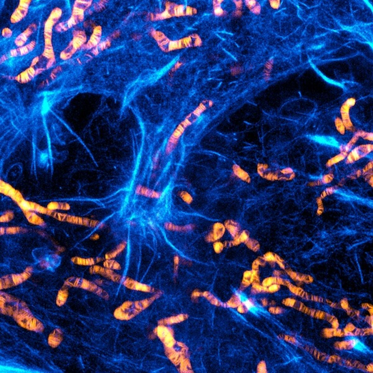
PKmito dyes are bright, photostable, non-phototoxic mitochondrial probes developed in the lab of Zhixing Chen at Peking University. PKmito dyes label mitochondria in live cells with very high specificity. The unique and unmatched feature of PKmito dyes is their extremely low phototoxicity, due to the presence of the intramolecular triplet quencher cyclooctatetraene (COT).
PKmito probes accumulate in the mitochondrial inner membrane (IM) and allow imaging of mitochondrial cristae structure and dynamics by confocal and STED superresolution microscopy.
Flipper-TR fluorescent probes for mechanobiology
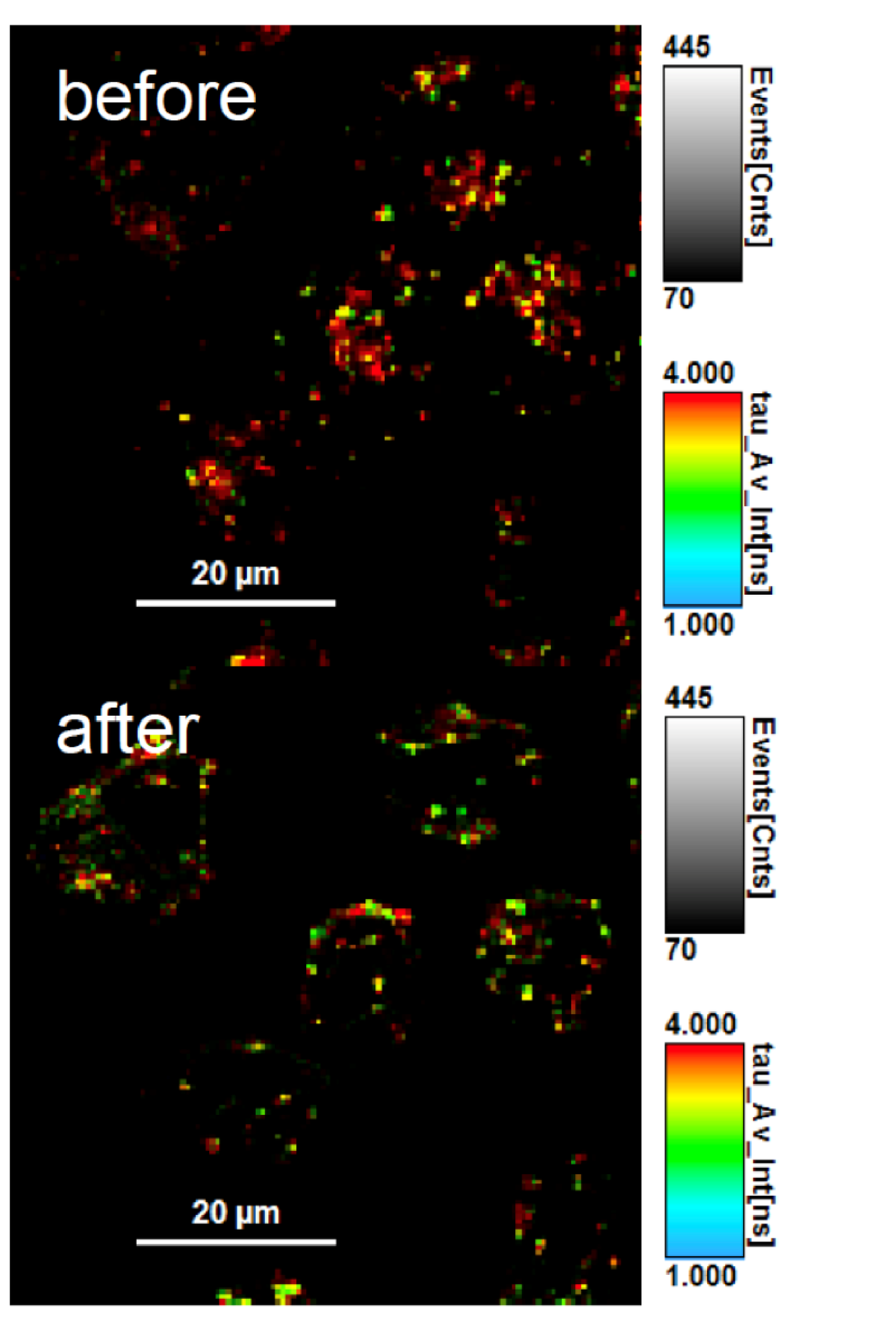
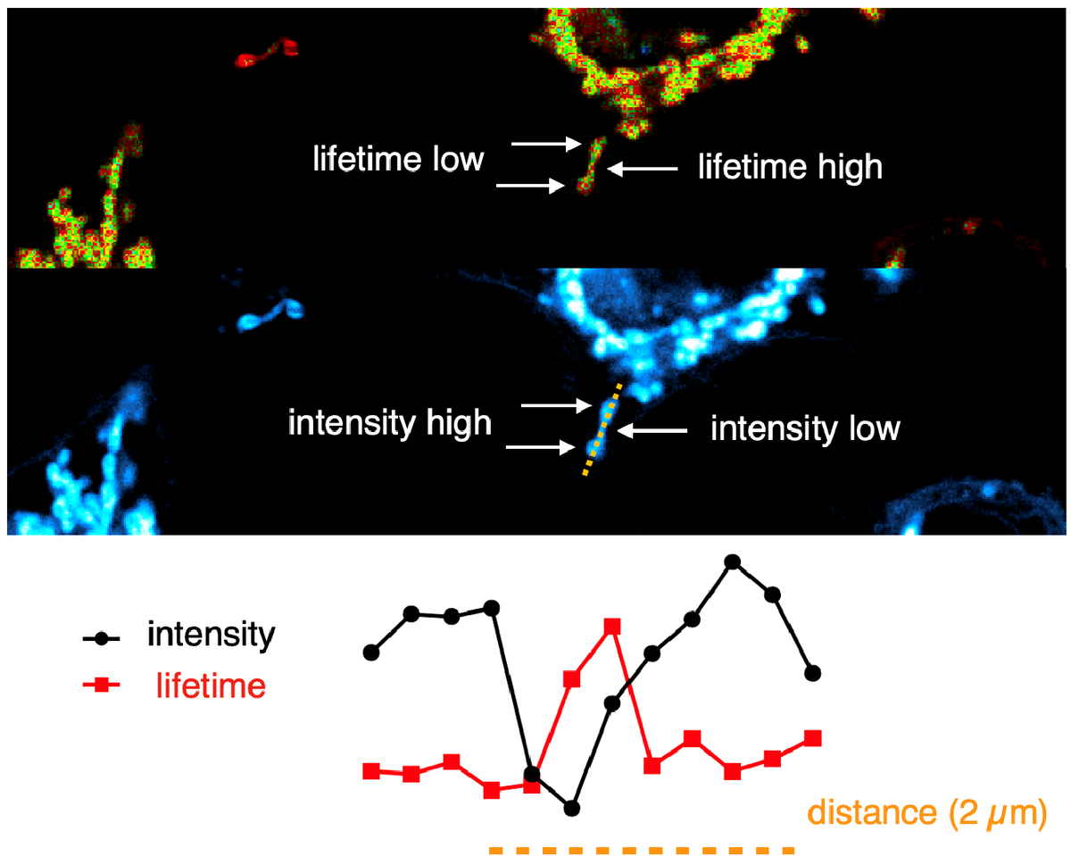
Flipper-TR® is a live cell fluorescent membrane tension probe. It is the first fluorescent membrane tension reporter developed for the field of mechanobiology. The fluorescence lifetime of Flipper-TR® is strongly dependent on the membrane tension, therefore through FLIM imaging it is possible to measure precisely the spatio-temporal distribution of tension in membranes. Flipper-TR® opens up a whole new field by giving access to real time membrane tension measurements in live cells.
SiR probes
Spirochrome’s SiR probes are based on the fluorphore Silicon Rhodamine (SiR). Its key features are:
- Cell permeability
- Fluorogenic character
- Compatibility with super-resolution microscopy
The fluorescence excitation and emission of SiR are in the far-red, reducing phototoxicity in live-cell imaging experiments. SiR is compatible with most microscopes as it can be used with standard Cy5 settings. The combination of all these properties set SiR-based probes apart from other fluorescent probes. Spirochrome SiR-probes work as simple as “add and image”
Spirochrome provides live cell imaging probes for DNA, Actin, Microtubules and lysosomes that are available in two colors: SiR and SiR700
SiR products
| Cat-No. | Item | Size | Price (CHF) |
|---|---|---|---|
| SC001 | SiR-actin kit | 50 nmol | 495.00 |
| SC002 | SiR-tubulin kit | 50 nmol | 495.00 |
| SC007 | SiR-DNA kit | 50 nmol | 330.00 |
| SC012 | SiR-Lysosome kit | 50 nmol | 365.00 |
| SC013 | SiR700-actin kit | 35 nmol | 495.00 |
| SC014 | SiR700-tubulin kit | 35 nmol | 495.00 |
| SC015 | SiR700-DNA kit | 35 nmol | 330.00 |
| SC016 | SiR700-lysosome kit | 35 nmol | 365.00 |
| SC604 | SiR700-BG | 35 nmol | 330.00 |
SiR fluorophore and reagents
Spirochrome provides SiR-reagents for the preparation protein, nucleic acid, peptide or small molecules SiR-conjugates creating tailor-made probes. Currently, carboxylic acid, NHS ester, maleimide, alkyne, azide, tetrazine and BCN derivatives of SiR are available.


