Founded by researchers from the Human Protein Atlas
Atlas Antibodies is the originalmanufacturer of highly validated primary antibodies andcontrol antigens. The company was founded by researchers from the prestigious Human Protein Atlas project who wanted to make the unique antibodies developed in the project available to fellow researchers worldwide.
The Primary Antibodies portfolio covers different research areas:
- Neuroscience
- Cancer
- Cell Biology
- Stem Cell
Atlas’ antibodies are developed against recombinant human Protein Epitope Signature Tags (PrESTs) of 50 to 150 amino acids and were designed to have the lowest possible identity to other human proteins. The specificity and purity are validated by protein array against 364 human recombinant protein fragments.
Atlas Antibodies believe in transparency. As a result, their antibody characterization data including images can be found on their website and the Human Protein Atlas. In addition, they specify the exact antigen sequence used to raise the antibody.
Product offering
For IHC, WB and ICC-IF applications
- Triple A polyclonal antibodies: rabbit polyclonals developed for the Human Protein Atlas, now available for researchers world-wide. Typically contain 3-5 epitopes. Ideal for IHC.
- PrecisA monoclonal antibodies: mouse monoclonals developed in-house, cultured in murine cell lines
- Blocking antigens
- Antibody panels (organelle markers, laminin markers, neuroscience markers, EMT markers)
- Loading control antibodies
What are the advantages of using Atlas Antibodies?
- They are made by researchers for researchers.
- Their results are reproducible: A unique purification process using the recombinant PrEST-antigen as the affinity ligand achieves the very highest level of reproducibility.
- Each Triple A Polyclonal antibody has been used for IHC-staining of samples from 708 individuals originating from normal and cancer afflicted human tissues.
- They have superior versatility across applications including IHC, IF and WB.
- All Triple A Polyclonals are presented on the Human Protein Atlas portal with extensive IHC characterization data. The Human Protein Atlas presents the complete human proteome, a map of all major tissues and organs in the human body.
Specificity & reproducibility
In order to guarantee specificity, Atlas Antibodies extensively validates and characterizes all their antibodies in the applications they have been approved for: IHC, ICC-IF and/or WB. In a second step (‘Enhanced Validation’, see video on the right), they use additional validation methods as proposed in the guidelines of the International Working Group for Antibody Validation (IWGAV) in Nature Methods.
As antibodies also need to be reproducible, each new lot is also compared to a previous lot in parallel, on a large number of samples and for each application the antibody is verified for.
Your specialist for IHC antibodies
Immunohistochemistry (IHC) is the most widely used application in histopathological diagnosis and research for the detection of proteins in tissues and cells. The use of antibodies to determine the presence and localization of specific proteins in surgical specimens is an indispensable tool in modern pathology.
How does Atlas Antibodies validate their IHC antibodies?
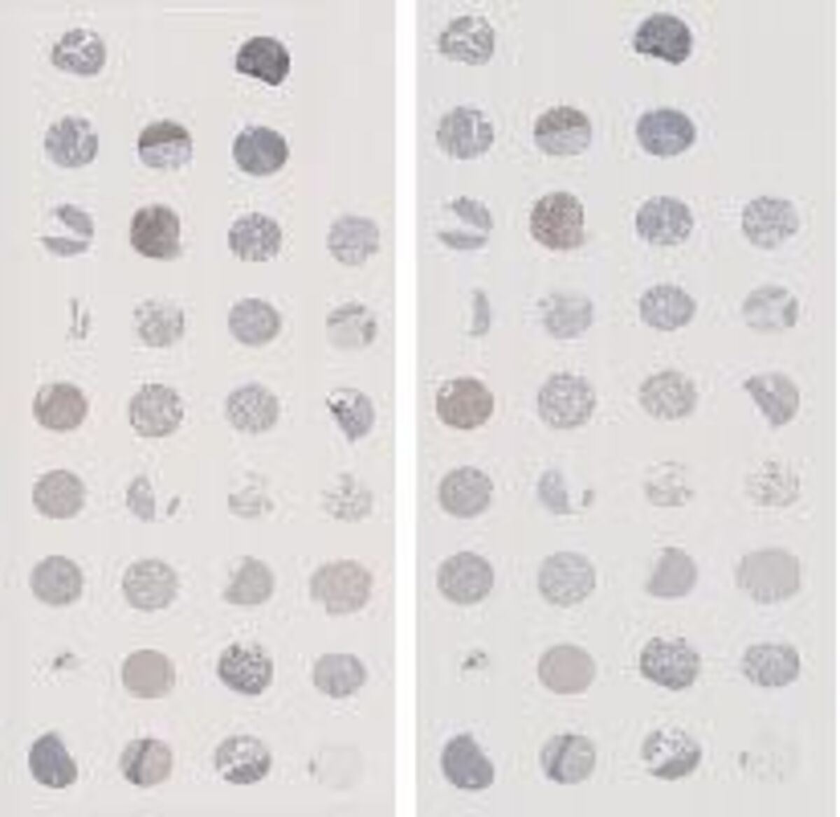
The antibodies verified for immunohistochemistry are characterized and validated by IHC-staining of formalin-fixed paraffin-embedded (FFPE) normal and cancer tissues in a tissue microarray format (TMA). As this is a non-quantitative method, experienced personnel perform thorough manual analysis of over 500 tissue cores for each antibody. They use normal and cancer tissues (high and low malignancy) from over 300 individuals.
In addition to this first round of validation, Atlas Antibodies further applies application-specific ‘Enhanced Validation’. For over 5’000 IHC-verified antibodies, at least one of the following two methods was used:
- Orthogonal validation: verification done with a non-antibody based method
- Independent antibodies validation: antibodies that bind to different parts of the protein are used, thus verifying each other
Orthogonal validation
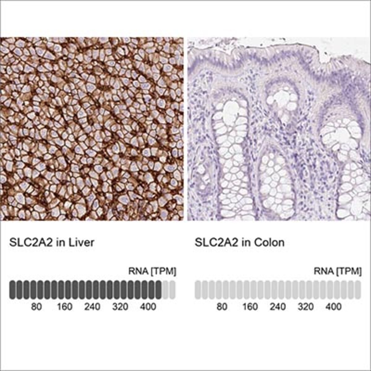
For orthogonal validation, the antibody staining is compared to RNA-Seq data for the same samples, using positive and negative samples. For an antibody to be validated, the IHC signal must match the RNA level. For each antibody, two tissues are chosen: one with a high RNA expression and one with no or low RNA expression. The difference in RNA expression must be at least fivefold.
Image: Example of orthogonal validation. IHC staining of liver and kidney tissues using the Anti-SLC2A2 antibody (HPA028997). The corresponding RNA-Seq data (TPM values) for the same tissues are presented below.
Independent antibodies validation
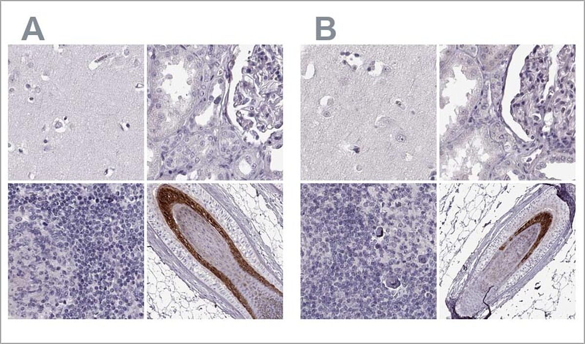
For this validation method, the staining patterns of at least two antibodies targeting the same protein but different, non-overlapping epitopes are compared. Over 20 tissues are evaluated for each antibody tested with this method.
Image: The two Anti-TCHHL1 antibodies HPA063483 (left, A) and HPA042579 (right, B) target different regions of TCHHL1. Antibody stainings across relevant positive and negative tissues are similar between the two, and the antibodies validate each other's staining pattern in IHC. Here, you can see IHC stainings of each of the two antibodies of cerebral cortex, kidney, lymph node and skin.
Are you interested in the validation methods used for WB and ICC-IF?
Read about them on Atlas’ website:
Innovative sustainable packaging alternative
Atlas Antibodies said goodbye to plastics and styrofoam, and transitioned to a fully sustainable, reusable, recyclable, and environmentally friendly option.
They have implemeted an eco-friendly solution by incorporating WoolCool (100% natural sheep wool) into our packaging. This change not only reflects Atlas' dedication to reducing their ecological footprint but also ensures that your Atlas Antibodies product is delivered with care for both you and the planet.


